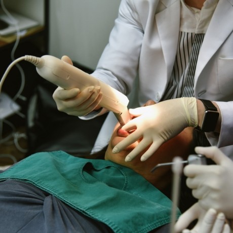
As a dentist who stays informed about the latest changes and advancements in dentistry, Dr. Mackie incorporates only the best, most innovative technologies and solutions into patients’ treatment plans. Whether it is the use of intraoral cameras to deliver more in-depth patient education, digital impressions for mess-free imaging, or The Wand™, which is used for more comfortable anesthesia delivery, you can expect greater efficiency, accuracy, and precision when it comes to achieving one-of-a-kind results.
Digital Impressions
Our iTero digital impression system eliminates the need for cold, messy dental putty. Instead of having you bite down and hold still to create a “decent” mold of your teeth, we can use our handheld device to scan your teeth and gums so that lab technicians can easily create a customized restoration. Whether it’s veneers, dental crowns, or dental bridges that you need, this unique system offers a mess-free, patient-friendly way to repair and improve smiles.
The Wand™
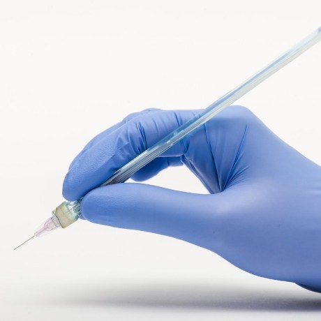
Many patients struggle with the idea of needles when visiting the dentist’s office. Since many of our services require the use of local anesthesia, it’s natural that we incorporate a more user-friendly way to administer this type of numbing agent. With The Wand™, we can control the speed at which the needle enters the soft tissues, creating a more comfortable experience that often leaves patients completely at ease and pain-free.
Intraoral Camera
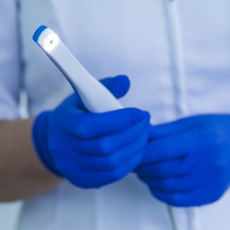
When looking to pinpoint even the smallest areas of decay, Dr. Mackie uses our SoproLife intraoral camera. This device projects real-time images of your teeth and gums onto a chairside monitor so that you can see what she sees. This not only increases the chances of catching problems early on by illuminating decay but also helps to keep patients informed about the importance of good oral hygiene.
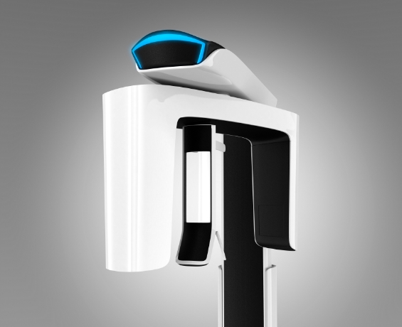
Digital X-Rays
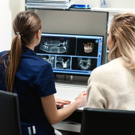
In the past, most dental professionals had to use traditional X-ray systems, which require film development. These days, we use digital X-rays due to their many benefits. A digital X-ray is safer and much quicker than its film predecessor. If the image comes out blurry, we can instantly retake a new one. Digital X-rays are a crucial diagnostic tool that helps us gain a better understanding of the inner workings of your mouth, more specifically the teeth, roots, and alveolar bone.
Why We Use Digital Radiography
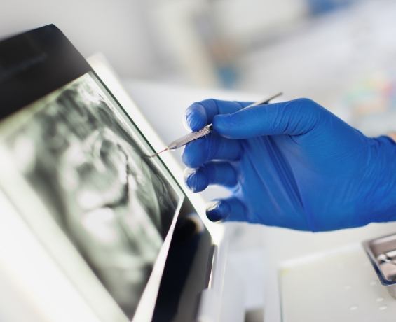
The use of digital dental radiography comes with many advantages. Unlike conventional film-based X-rays, digital radiographs allow us and you to immediately view dental images. This is extremely useful, especially when we perform surgical procedures such as dental implants.
Digital radiographs have enhanced images. We can magnify images for easier reading and precise analysis of manifesting dental issues. Also, because of their precision, digital radiographs do not require retakes due to failed focus on details. This protects our patients from extended radiation exposure. Overall, digital radiology is much safer for patients, emitting up to 80% less radiation than film-based radiology.
With digital radiology, we can easily store data in computers and other digital devices for future reference, consult with our other experts. We believe that the advantages of using digital radiology outweigh the high cost of equipment. Digital radiography equipment defines the two major types of digital radiography.
We believe that the advantages of using digital radiology outweigh the high cost of equipment. Digital radiography equipment defines the two major types of digital radiography.
Types Of Digital Radiography
Digital radiography uses sensors to capture images of your teeth and other oral structures. The types of radiography are defined based on the position of the sensor, either intraoral or extraoral radiographs.
When we do intraoral radiographs, we place the sensor inside the patient’s mouth to focus on a certain area of the dental structure. Intraoral radiographs can be Bitewings when the patient bites the sensor to allow our specialist to capture images of their crowns and notice dental caries. They can also be Periapicals when they target, we use these radiographs to determine the invisible causes of your tooth pain. Occlusals are radiographs that target the skeletal anatomy of your palate or the floor of your mouth and are excellent in noticing oral anomalies such as abnormally placed teeth. We use specific equipment for intraoral radiographs including digital sensors, and digital X-ray units.
With extraoral radiographs, we use the sensor outside your mouth to generate whole images of your dental structures. The digital image machine rotates around your head to create a comprehensive image. Extraoral radiographs include panoramic radiographs which show a curved image of your jaws and are optimal in revealing the overall status of your dentition and surrounding structures.